Evaluation of reduction quality and implant positioning in intertrochanteric fracture fixation: A review of key radiographic parameters
Authors: Matthias Wittauer, Joseph Henry, Guillermo Sánchez-Rosenberg, Anton Philip Lambers, Christopher W Jones, Piers J Yates
Site: Fiona Stanley Hospital, WA
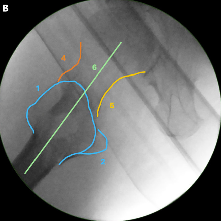
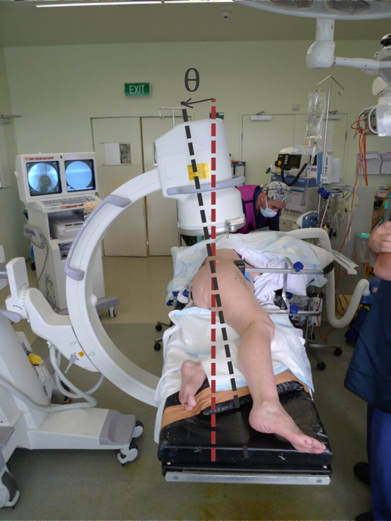
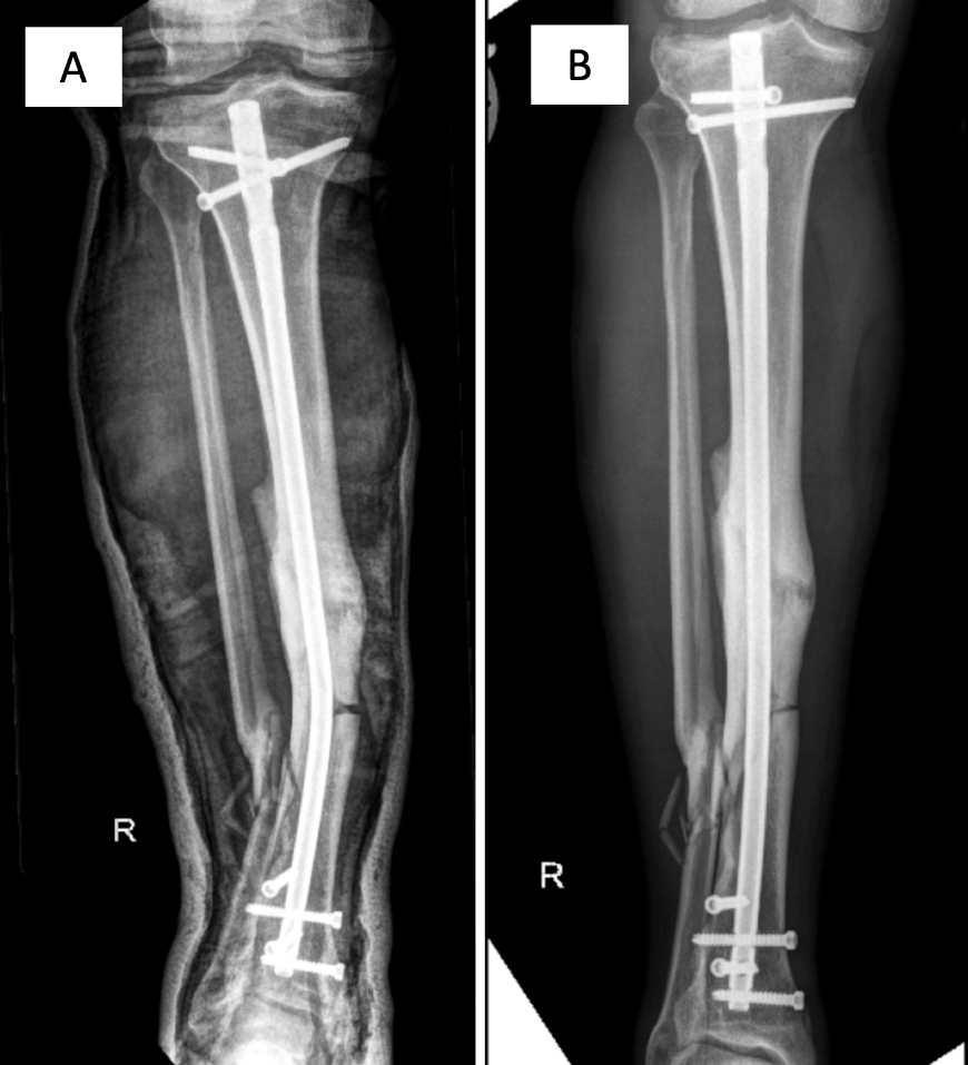
This article, co-authored by Dr Anton Lambers, reviews the most important X-ray measurements surgeons use when repairing a common type of hip fracture in older adults, called an intertrochanteric fracture. These injuries can be life-threatening and often require surgery using a metal rod or screw. The review explains how getting the broken bone ends lined up accurately (“reduction”) and placing the implant in the best position are vital for a strong repair and to avoid complications that can require further surgery. It outlines simple but reliable ways surgeons can check their work during the operation, such as specific angles, lines, and distances visible on X-rays, to help ensure the bone heals in the correct shape and the implant remains secure.
As a contribution to the medical literature, this paper brings together the most up-to-date research into one clear guide for orthopaedic surgeons worldwide. By summarising the key radiographic checks—like tip-apex distance, cortical support, and reduction quality scores—it provides a practical reference that can improve surgical precision and patient outcomes. For patients, the takeaway is reassuring: following these principles helps surgeons fix hip fractures more accurately, lowering the risk of mechanical failure, reducing pain, and supporting a quicker return to mobility.
